Cavum septum pellucidum closure to form a single septum pellucidum occurs by 6 months of age in 85% of cases but may persist to adulthood in 15% 3. Because of this ordered obliteration, a cavum septum pellucidum almost always accompanies a cavum vergae and is referred to as cavum septum pellucidum et vergae 1,2. History and etymology. Cavum septum pellucidum (CSP) cyst, cavum vergae, and cavum velum interpositum are various presentations of benign midline anterior intracranial cysts. They are pathological when symptomatic, which arise depending upon the size of the cysts. Cavum septum pellucidum cysts are rare lesions with an incidence of 0.04% . Symptomatic cysts of CSP are.

🥇 Anatomía del SEPTUM PELLUCIDUM. ¡Explicación Rápida y Sencilla! YouTube

Cavum septi pellucidi and cavum vergae MedLink Neurology

Absent Cavum Septi Pellucidi American Journal of Obstetrics & Gynecology

Cavum septi pellucidi Ars Neurochirurgica

Cavum septi pellucidi and cavum vergae MedLink Neurology
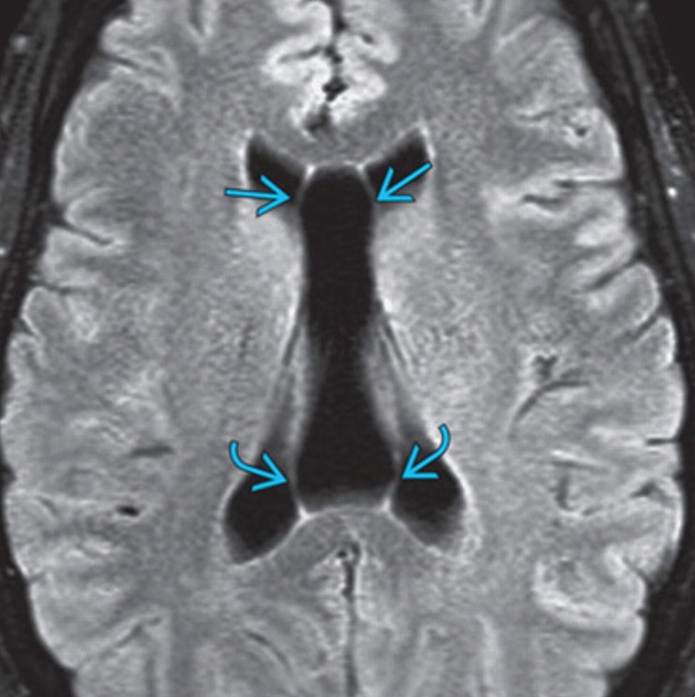
Radiology case Cavum septi pellucidi, cavum vergae

Cavum Septi Pellucidi (CSP) Brain imaging

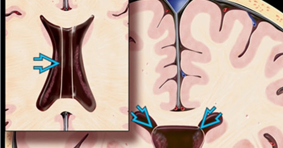
Cavum septum pellucidum, cavum vergae, and cavum veli interpositi (annotated CT) Image

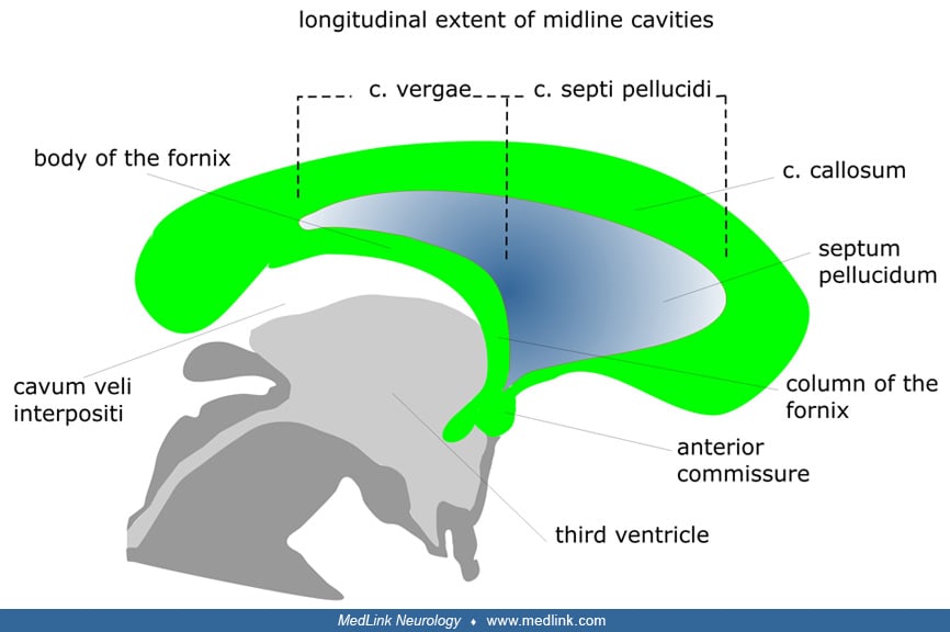
Lateral and frontal line diagrams of the cavum septi pellucidi (green)… Download Scientific

Abnormalities Associated With the Cavum Septi Pellucidi on Fetal MRI What Radiologists Need to
Cavum Septi Pellucidi (CSP) Brain imaging
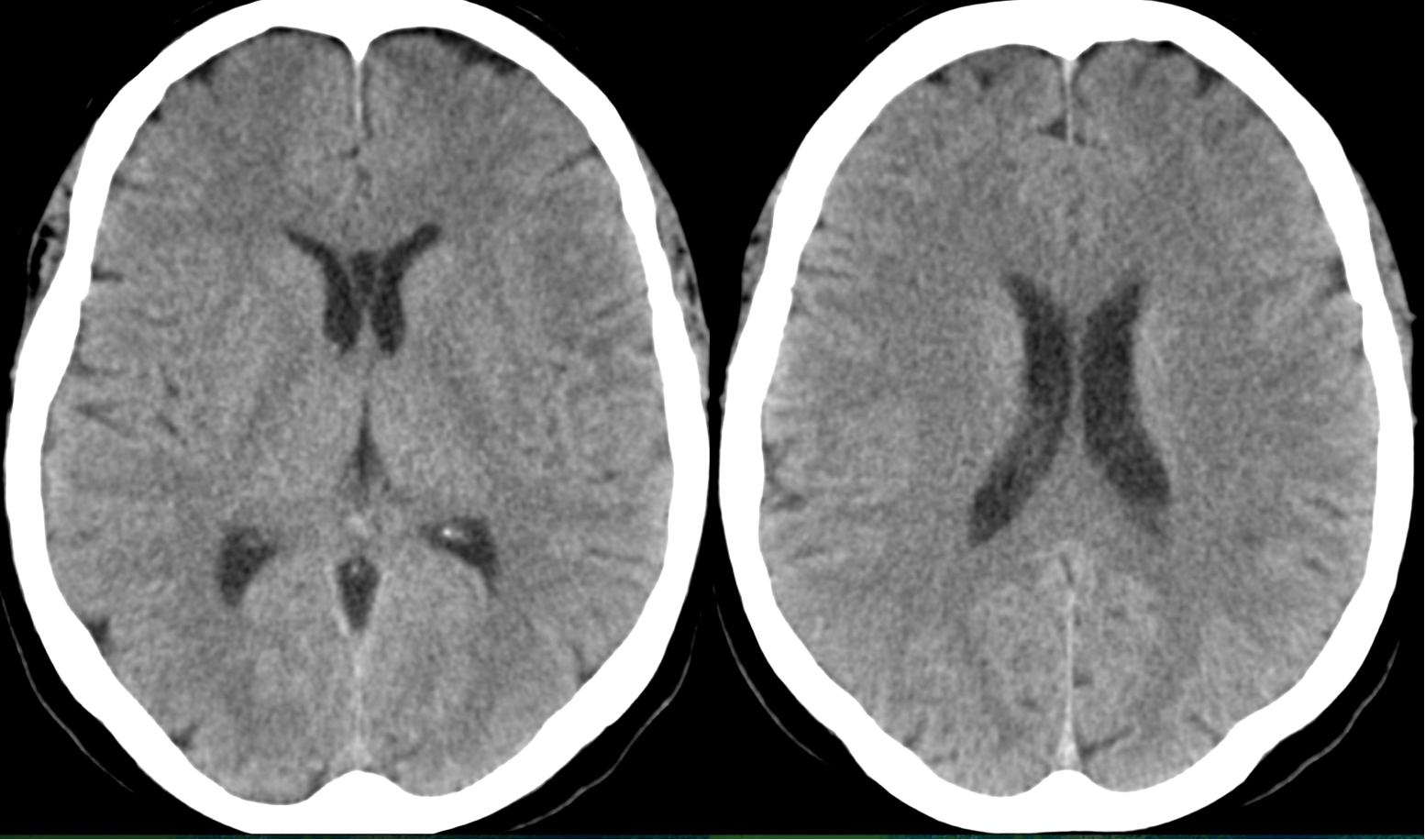
Cavum septum pellucidum, cavum vergae, and cavum veli interpositi (annotated CT) Image

Cavum septum pellucidumMRI Sumer’s Radiology Blog

Cavum Septum Pellucidum Diagram

Abnormalities Associated With the Cavum Septi Pellucidi on Fetal MRI What Radiologists Need to

Abnormalities Associated With the Cavum Septi Pellucidi on Fetal MRI What Radiologists Need to
Cavum Septi Pellucidi (CSP) Brain imaging

Abnormalities Associated With the Cavum Septi Pellucidi on Fetal MRI What Radiologists Need to
Cavum septi pellucidi and cavum vergae MedLink Neurology

Cavum Vergae
Cavum septi pellucidi and cavum vergae in normal and developmentally delayed populations.. 12. Ho YK, Turley M, Marc-Aurele KL, Jones MC, Housman E, Engelkemier D, et al.. Enlarged cavum septi pellucidi and vergae in the fetus: a cause for concern. J Ultrasound Med 2017; 36: 1657-68. doi:.. Background Cavum septum pellucidum (CSP) and cavum vergae (CV) are normal anatomical variations present in some children, adolescents, and adults. When the cavity is larger than normal, it is.


